6.5 - Medical Imaging
Xray production
Electrons are emitted into a vacuum tube via thermionic emission. An external power supply produces a massive p.d. between the anode and cathode, causing the electrons to be rapidly accelerated in this high-voltage electric field. Then they are rapidly decelerated with collisions with a hard metal anode, causing them to lose KE (~1%) which is emitted as xrays. Rest is lost to the anode as thermal energy. Their KE is transformed into high-frequency photons of EM radiation. This radiation is called Bremsstrahlung radiation.
Xray focusing
Straight, parallel xrays are created by directing xrays to a thin window which enters a collimator that absorbs xrays that are not parallel to it. Xray energy is absorbed by tissue as it passes through the body, and how effective this absorption is depends on the attenuation coefficient mu, which is constant for different materials. The energy before entering and after exiting the body can be measured. These differences can be visualised on photographic film or a digital image to view the targeted body part.
Xray attenuation
"Exponential decay"-like formula for xray intensity loss:
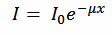
- I = Intensity
- I0 = Initial intensity
- Mu = Attenuation coefficient
- x = Distance traveled through body
Mu α z^3
... where z is the proton number of the material.
Attenuation mechanisms
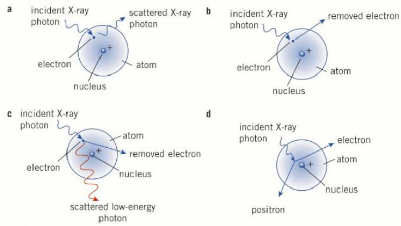
Simple Scattering (a)
Simple scattering is when xray photons reflect off of bone if they do not have sufficient energy to do more complex scattering.
Photoelectric Effect (b)
Via the photoelectric effect, xray photons are absorbed by electrons into the material, releasing photoelectrons.
Compton effect (c)
In the Compton effect, a photon interacts inelastically with an electron. The photon transfers some of its energy and momentum to the electron. The photon is scattered with less energy, while the electron is removed from the atom. Both energy and momentum are conserved in this interaction.
Pair Production (d)
Pair production is when an electron-positron pair is spontaneously created as the xray passes through the electric field of an atom. The required energy is about 1.02MeV, as derived below:
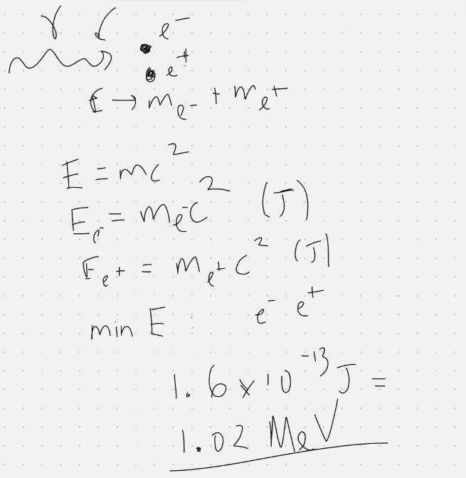
Not very applicable in medical imaging, as xrays usually don't have this much energy.
Medical Tracers
Medical tracers are substances injected into a body. Their position in the body can be detected.
- Fluorine-18 for beta+ decay. Protons decay into neutrons, releasing a positron and an electron-neutrino. The positron annihilates with an electron, releasing two gamma photons travelling in exactly opposite directions which are detected.
- Technetium-99m decays to immediately release gamma photons. Primarily used to monitor major organs. The "m" means metastable, meaning it remains in a high energy state for prolonged periods of time, eventually decaying to a far more stable isotope of technetium.
Gamma detection
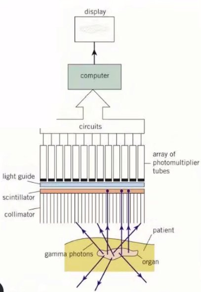
- Gamma photons pass through the collimator if their direction of travel is parallel to it. Otherwise, they are absorbed by it.
- These high-energy photons interact with the scintillator, releasing multiple more lower energy photons.
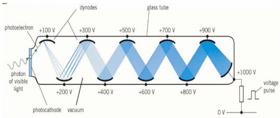
- The light guide/photocathode absorbs the photons and releases photoelectrons via the photoelectric effect.
- These electrons travel through the photomultiplier tube, generating more electrons to be passed into a computer for imaging.
In the photomultiplier tube, a photoelectron hits the first plate, releasing more electrons that hit the next plate, which causes more electrons to be released to hit the next plate. This repeats, generating lots of electrons that can create a substantial current.
CAT Scans
Uses xrays to create a 3-dimensional image of a person's body.
The machine is a ring that the person's body passes through. It generates a fan shaped beam of xrays to take 2d slices of the person's body, which are picked up by a ring of elecronic detectors. The images are stacked on top of each other in a computer program to create the 3D product.
The resolution of the image is greater and can distinguish between different soft tissues, which 2D xrays cannot do.
However, it takes significantly longer than a 2D xray, so the patient is exposed to a higher dose of ionising radiation. About 10-30 minutes.
PET Scans
PET scans detect gamma rays generated from annihilation between positrons and electrons to create a 2D or 3D image. It has a ring of gamma detectors/cameras that pass up and down the patient to generate a 3D image.
Positron-electron annihilation releases two photons in opposite directions. The ring can pick up both of these and use the time delay between their arrivals to calculate the exact location of the annihilation event, characterising the depth inside the body. This means PET scans have a higher resolution than other gamma cameras.
Ultrasound
(A piezoelectric material generates a voltage when it is contracted or expanded, or will contract and expand when a voltage is applied.) Applying aan alternating voltage to a piezoelectric crystal cancauses produceit to contract and expand at the same frequency as the alternating source, producing ultrasound vibrations, and a crystal receiving them can generate an alternating voltage.waves.
An ultrasound transducer has an alternating potential difference causing repetitive compression and stretching of the crystal. The crystal's resonant frequency is chosen to increase intensity. After creation of the ultrasound, the potential difference is removed and the reflected signal is read.
- An A type ultrasound scan produces a very low resolution 1D image. It is used to determine distances from the ultrasound device to the point of reflection. This is achieved by measuring time delay between generating and receiving the signal, using the speed of sound to approximate distance.
- A B type ultrasound scan produces a 2D image by moving the transducer over the patient's skin. It is effectively a series of A type scans stitched together to form an image.

... where Z is acoustic impendence,impedance, rho is the density, and c is the speed of sound in the material.

Ir is the intensity of reflected ultrasound. I0 is the intensity of incoming ultrasound. The ratio is known as the reflection coefficient.
A gel is put on the skin before an ultrasound. It has a similar acoustic impendenceimpedance to skin to minimise the amount of reflected ultrasound waves.
The Doppler Effect
The Doppler effect is the observed change in the frequency of a wave when it is reflected off of or produced by a moving source, e.g. an ambulance siren.

... where delta f is the observed shift in frequency, f is the original frequency, v is the speed of blood flow, theta is the angle between the probe and blood flow direction, and c is the speed of ultrasound in blood.